The cauda equina is the group of bundled nerves which branch off the bottom of the spinal cord forming a mass of nerve roots in the central canal space which continue to exit at each vertebral level of the spine. The spinal cord ends at individually varying locations between the T12 and L3 vertebrae in normal adults. After the end of the cord, the spinal nerve roots continue downwards through the lumbar, sacral and coccyx regions to serve the neurological needs of the lower body and lower extremities. The name of the structure translates into horse’s tail, which is very appropriate. The tail-shape of the nerve mass gradually thins from a wide grouping of nerves to a single nerve exiting the center of the vertebral column at the final coccyx level.
This discussion helps to educate patients about the importance of the bundled spinal nerves and how these tissues might be involved in a painful dorsalgia condition.
Cauda Equina Facts
The spinal nerve roots branch off from the spinal cord and continue through the central canal exiting between the vertebrae at their designated levels. However, there is no spinal cord after the upper lumbar region, so the horse’s tail takes its place as the central neurological structure. These are the nerve roots most often implicated in pinched nerve conditions in the lumbar spine, since they can be directly affected by both foraminal and central canal stenosis.
The nerves in this region of the back are often blamed for many varied painful syndromes including common lower back pain and sciatica. These nerves are also extremely susceptible to oxygen deprivation from a regional process, since they are all bundled in a small area and can be affected en masse.

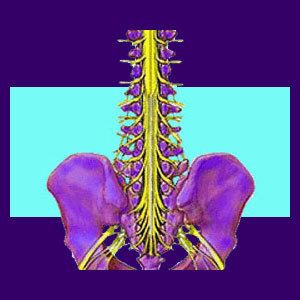
Structural Anatomy
The nerve roots branch off the spinal cord where the cord ends, usually at L1 or L2. The nerves descend together through the remainder of the spinal vertebrae, giving the appearance of a horse’s tail, hence the name of the structure. As each nerve reaches its designated level, it exits from the spinal column to supply the network of nerves serving a particular anatomical region.
This grouping of neurological tissue gradually thins as the individual nerves leave the spine, creating a wedge shape which tapers from wider to thinner, until only a single coccygeal nerve root remains.
Nerve Compression in the Horse’s Tail
Pinched or compressed nerves in this region can create a medical emergency for the patient. This condition is known as cauda equina syndrome and usually requires immediate surgical correction in order to restore neurological function and prevent permanent nerve damage. This condition is usually the result of massive trauma to the lower spine and can leave lasting effects even if corrected immediately.
Common symptoms include numbness in the buttocks and genitals, sexual dysfunction, paraplegia, incontinence and loss of bowel control. This condition is extremely serious and should be treated by a qualified hospital emergency unit immediately.